Cross Section Of A Bone / A and P Lab section 8 Flashcards | Quizlet
Get link
Facebook
X
Pinterest
Email
Other Apps
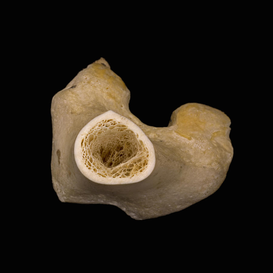
Cross Section Of A Bone / A and P Lab section 8 Flashcards | Quizlet. Related posts of cross section of a long bone bone test anatomy and physiology. Bone is a specialised type of connective tissue. Start studying cross section of bone. Sections of bone marrow tissue. At the outer regions of the section, you can see a dense, thick layer of compact bone.
Muscle attachments are visible along the outer surface. The outlined area is a cross section of an osteon of compact bone. And why does the marrow stop where it does, and so sharply? There are three general classes of bone. The central tubular region of the bone, called the diaphysis, flares outward near the end to form the metaphysis, which contains a largely cancellous, or spongy, interior.
Bone Cross Section, Lm Photograph by Science Stock Photography from images.fineartamerica.com The upper (biting) surfaces of the tooth are at top, with the lower sections (bottom) embedded in the gums and jaw bone (not shown). Bone markings the surface features of bones vary considerably, depending on the function and location in the body. An outer 'fibrous layer' containing mainly fibroblasts, and an inner 'cambium layer' containing progenitor cells. Each bone in your body is made up of three main types of bone material: The surface features of bones vary considerably, depending on the function and location in the body. Two types of bone tissues in cross section of a long bone : The cell line involved in osteogenesis consists of preosteoblasts, osteoblasts, osteocytes and bone. Learn vocabulary, terms, and more with flashcards, games, and other study tools.
There are three general classes of bone.
There are three general classes of bone. The arrows point toward the tumor. Bone in arm pictures 12 photos of the bone in arm pictures bone cancer arm pictures, pictures of bone cancer in arm, bone, bone cancer arm pictures, pictures of bone cancer in arm. In this short video i use blender 2.8. It consists of two layers; The upper (biting) surfaces of the tooth are at top, with the lower sections (bottom) embedded in the gums and jaw bone (not shown). The compact bone is made up of osteon. Bone is a specialised type of connective tissue. Cross section of mandible at first molar region showing cortical and spongy bone basic concepts in osteogenesis. Cross section of a bone. In the center of each osteon is the central canal, a space that houses blood vessels and nerves that supply bone. Compact bone is the outer layer and the spongy bone forms the inner layer. There are trabeculae in spongy bone which gives its sponge like appearance.
While it is not as hard as compact bone, spongy bone plays an important role of protecting the marrow where blood cells are produced. Start studying cross section of a long bone. It has a unique histological appearance, which enables it to carry out its numerous functions: Compact bone, spongy bone, and bone marrow. An outer 'fibrous layer' containing mainly fibroblasts, and an inner 'cambium layer' containing progenitor cells.
Project Beak: Adaptations: Skeletal System: Hollow Bones from projectbeak.org Bone is a dynamic biological tissue, composed of various metabolically active cells that are integrated into a rigid framework. Related posts of cross section of a long bone bone test anatomy and physiology. This is known as the periosteum. An outer 'fibrous layer' containing mainly fibroblasts, and an inner 'cambium layer' containing progenitor cells. Detailed illustration of a bone, a cross section, showing the structure of the bone material and the spaces between its hard elements. Bone cross section view : Compact bone is the outer layer and the spongy bone forms the inner layer. Thanks to a special coloring process, the mini joint series features extremely realistic bones.
The inner portion of the bone is composed of trabecular bone and the intervening bone marrow.
Start studying cross section of a long bone. Learn vocabulary, terms, and more with flashcards, games, and other study tools. At the outer regions of the section, you can see a dense, thick layer of compact bone. There are trabeculae in spongy bone which gives its sponge like appearance. Table 1 describes the bone markings, which are illustrated in (figure 4). Bone cross section view : In three dimensions an osteon is cylindrical in shape. I don't find it enhances the image. The compact bone is made up of osteon. Concentric layers of bone cells (osteocytes) and bone matrix surround the central canal. As the names suggest compact bone looks compact and the spongy bone looks like sponges. Posteriorly the body is connected to a thin ring of bone known as the arch. In the center of each osteon is the central canal, a space that houses blood vessels and nerves that supply bone.
There are three general classes of bone. I don't find it enhances the image. The upper (biting) surfaces of the tooth are at top, with the lower sections (bottom) embedded in the gums and jaw bone (not shown). Bone is a specialised type of connective tissue. From wikimedia commons, the free media repository.
Treatment of Avascular Necrosis of the Femoral Head with ... from www.itmonline.org Muscle attachments are visible along the outer surface. Bone cross section view : Concentric layers of bone cells (osteocytes) and bone matrix surround the central canal. Table 1 describes the bone markings, which are illustrated in (figure 4). Would it be a good thing to show the epiphyseal plate? Each bone in your body is made up of three main types of bone material: Learn vocabulary, terms, and more with flashcards, games, and other study tools. There are three general classes of bone.
Would it be a good thing to show the epiphyseal plate?
In the center of each osteon is the central canal, a space that houses blood vessels and nerves that supply bone. As the names suggest compact bone looks compact and the spongy bone looks like sponges. Table 1 describes the bone markings, which are illustrated in (figure 4). This is known as the periosteum. And why does the marrow stop where it does, and so sharply? The compact bone is made up of osteon. Marrow in the shaft of long bones is typically yellow, with red marrow in the head through the cancellous bone. Compact bone is the outer layer and the spongy bone forms the inner layer. Concentric layers of bone cells (osteocytes) and bone matrix surround the central canal. At the outer regions of the section, you can see a dense, thick layer of compact bone. The inner portion of the bone is composed of trabecular bone and the intervening bone marrow. Photomechanical print page item number: Why is the marrow red?


Comments
Post a Comment